
dissected frog Stock Photo Alamy
Frog Internal Anatomy - Dissection Guide. Lay the frog on its back, spread out its limbs, and pin them to the tray. Use forceps to lift the skin between the hind legs and make a small incision with a scalpel. Continue the cut up the center of the frog's body with scissors, being careful to cut through the skin only.
13 Frog Dissection Worksheet /
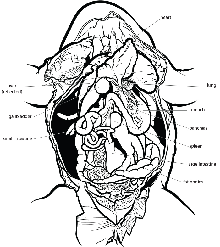
Frog Dissection, Labeled Images Frog Dissection Resources Frog Dissection: Complete Guide - includes external anatomy, mouth, and the organs of the abdominal cavity, download available in pdf and google doc. Frog Dissection Overview - introduction to the lab, Google Slides Frog External Anatomy - legs, eyes, mouth structures
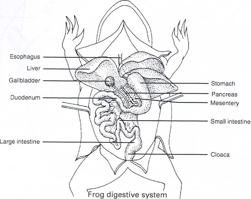
Frog Dissection Diagram and Labeling
diagram of a dissected frog of class 9 can be tricky for some people in this video I will show steps to draw a dissected frog.If you like my effort don't fo.

Anatomy for dissected body frog diagram Royalty Free Vector
Frog dissection is a process of examining the internal anatomy of a frog by making cuts through the skin and muscles. During the dissection, students typically examine the organs, including the heart, lungs, liver, stomach, and intestines. In fact, this is quite a sensitive process and requires some specific tools.

Frog Anatomy Coloring Worksheet Biology LibreTexts
5. First, cut the length of the frog, then cut across the frog horizontally just under the arms and at the top of the thighs. Be careful that you cut muscle. Avoid cutting into the organs of the frog. The trick to this is to go slowly, revealing a little at a time.
puppies123 Frog Dissection
Dissection Instructions. Place the frog in the dissecting pan ventral side up. Use scissors to lift the abdominal muscles away from the body cavity. Cut along the midline of the body to the forelimbs. Make transverse (horizontal) cuts near the arms and legs. Life the flaps of the body wall and pin back.
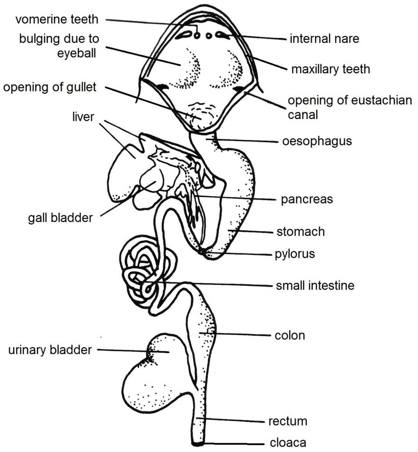
Digestive system of frog Anatomy and Physiology of digestion Online
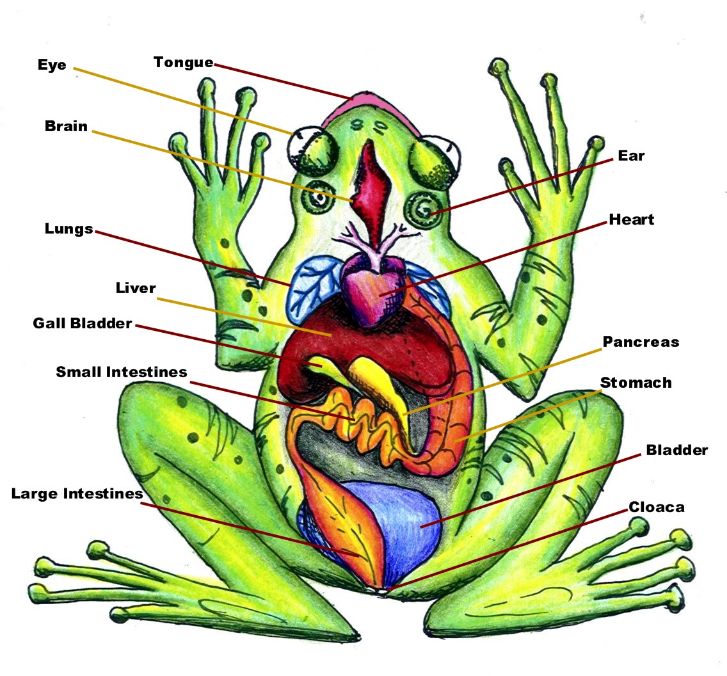
Frog Dissection Frogs are excellent model organisms for scientific studies of development, behavior, anatomy, and physiology. They are commonly used in biology classes as representative vertebrates with specialized amphibian characteristics and behaviors.

dissected frog Frog dissection, Frog, Diagram
Background: As members of the class Amphibia, frogs may live some of their adult lives on land, but they must return to water to reproduce. Eggs are laid and fertilized in water. On the outside of the frog's head are two external nares, or nostrils; two tympani, or eardrums; and two eyes, each of which has three lids.
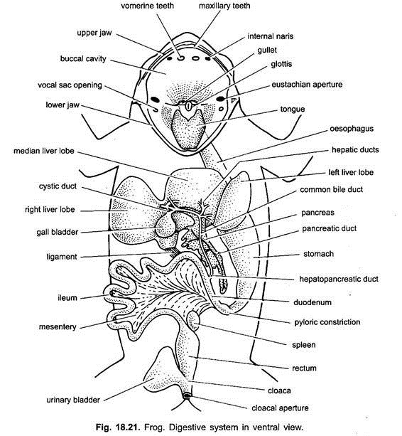
Digestive System of Frog (With Diagram) Vertebrates Chordata Zoology
1. Place the frog in the dissecting pan ventral side up. 2. Use scissors to lift the abdominal muscles away from the body cavity. Cut along the midline of the body to the forelimbs. 3. Make transverse (horizontal) cuts near the arms and legs. 4. Life the flaps of the body wall and pin back.

Frog anatomy educational vector illustration diagram Childrens
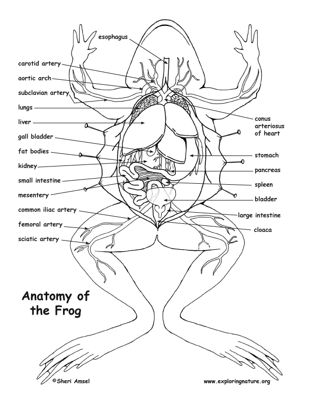
January 6, 2024 < http://www.exploringnature.org/db/view/Frog-Dissection-Diagram-and-Labeling > Frog Dissection Diagram and Labeling
Frog Dissection External Anatomy
Frog dissection is a common laboratory technique used to study the anatomy of the amphibian. The procedure involves opening up the frog's body and examining its internal organs, skeletal system, and circulatory system.

Arterial System of Frog Diagram Quizlet
Label the diagram of the external anatomy of a frog's body: (Word bank: head, trunk, forelimb, upper arm, wrist, hand, finger, digits, male pad, hindlimb, thigh, shank, ankle, foot, webbed toes). 1 2 9 6 4 8 5 7 3 11 12 13 10 14 15 2.
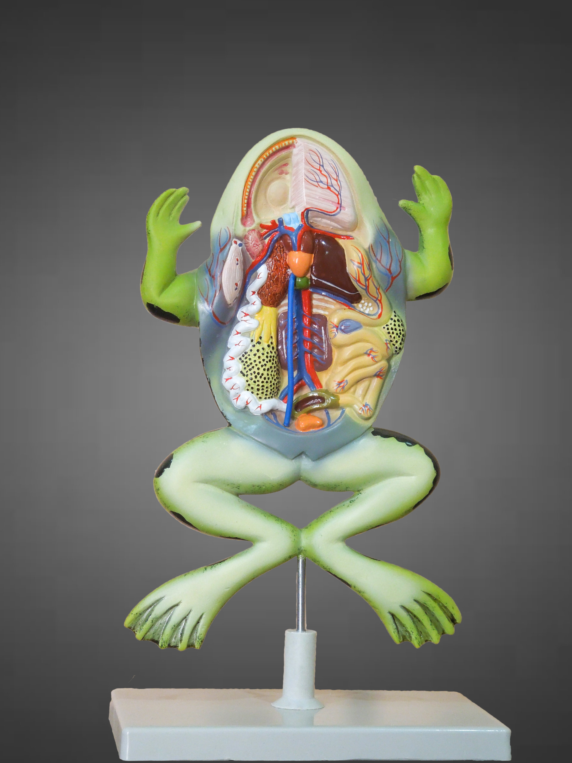
Frog Dissection Model Medilab Exports Consortium
0:00 / 4:06 How to draw Dissection of frog step 4 OPAL art 5.89K subscribers Subscribe 2.7K views 1 year ago 10th Biology Federal Board FSc, Biology practical copy, Experiment 25.
Frog Dissection MRS. MERRITT'S BIOLOGY CLASS
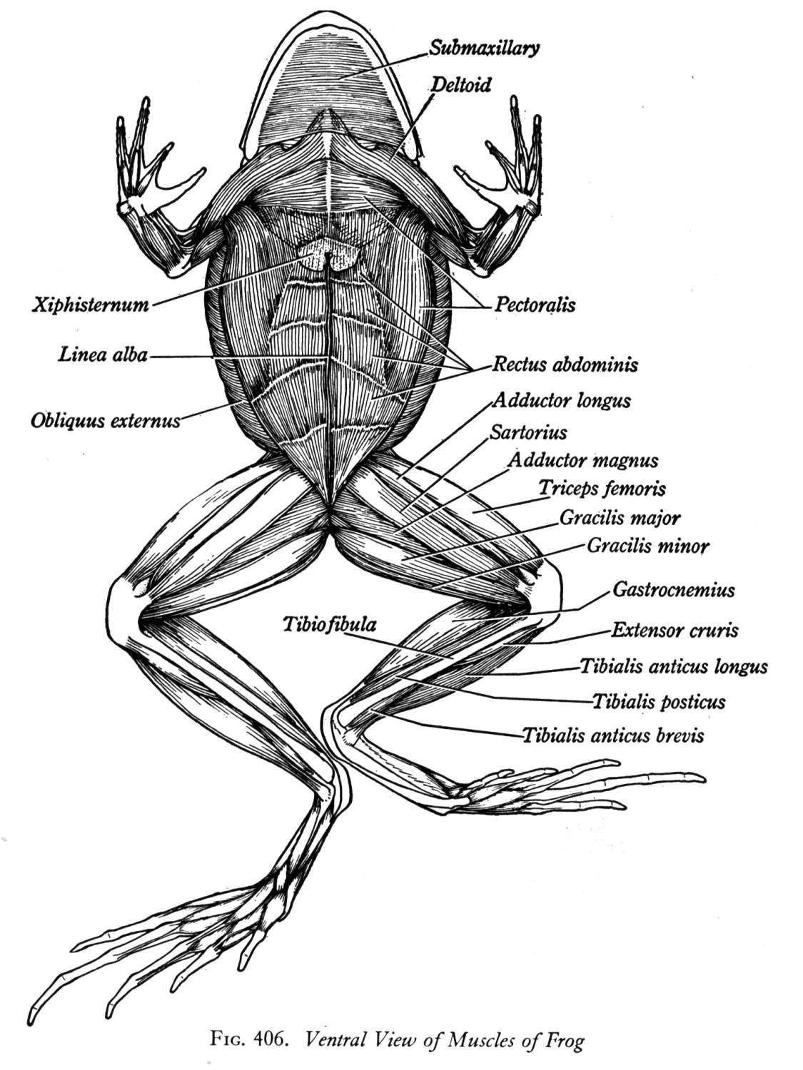
Frog Anatomy and Dissection Frog Dissection (2) Frog Dissection Alternative. Head and Mouth Structures. Vomerine Teeth: Used for holding prey, located at the roof of the mouth Maxillary Teeth: Used for holding prey, located around the edge of the mouth. Internal Nares (nostrils) breathing, connect to lungs. Eustachian Tubes: equalize pressure in inner ear.
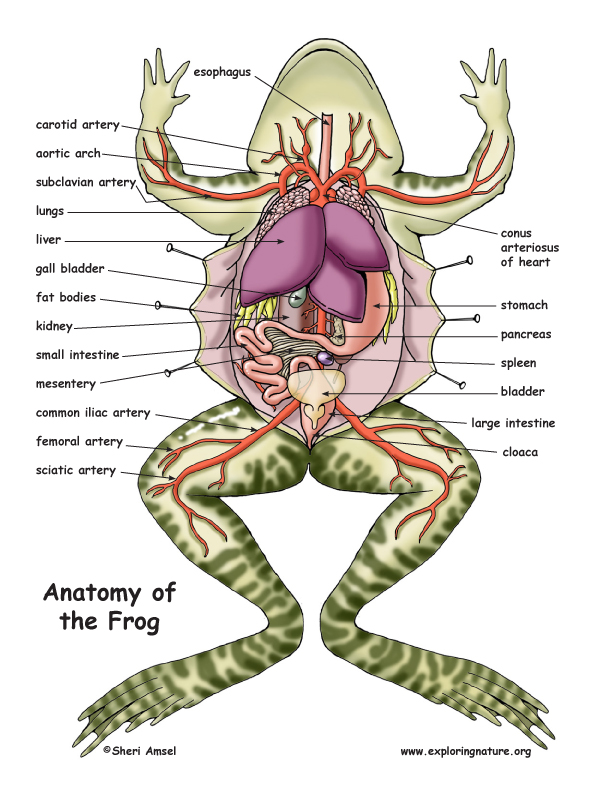
Frog Dissection Diagram and Labeling
Purpose: To locate,observe,and diagram the external (outside) structures of frogs. To see similarities between this organism and ourselves. Vocabulary: Dorsal-the back of an organism Ventral- the.
Frog Dissection
Bring 2 diagrams showing the internal anatomy of a frog. Describe each HUMAN system (you will later compare the humans systems to the frog after the dissection). 1. Human Circulatory System: a) Heart: o Total number of chambers ___________ o Number of atria__________ o Number of ventricles____________